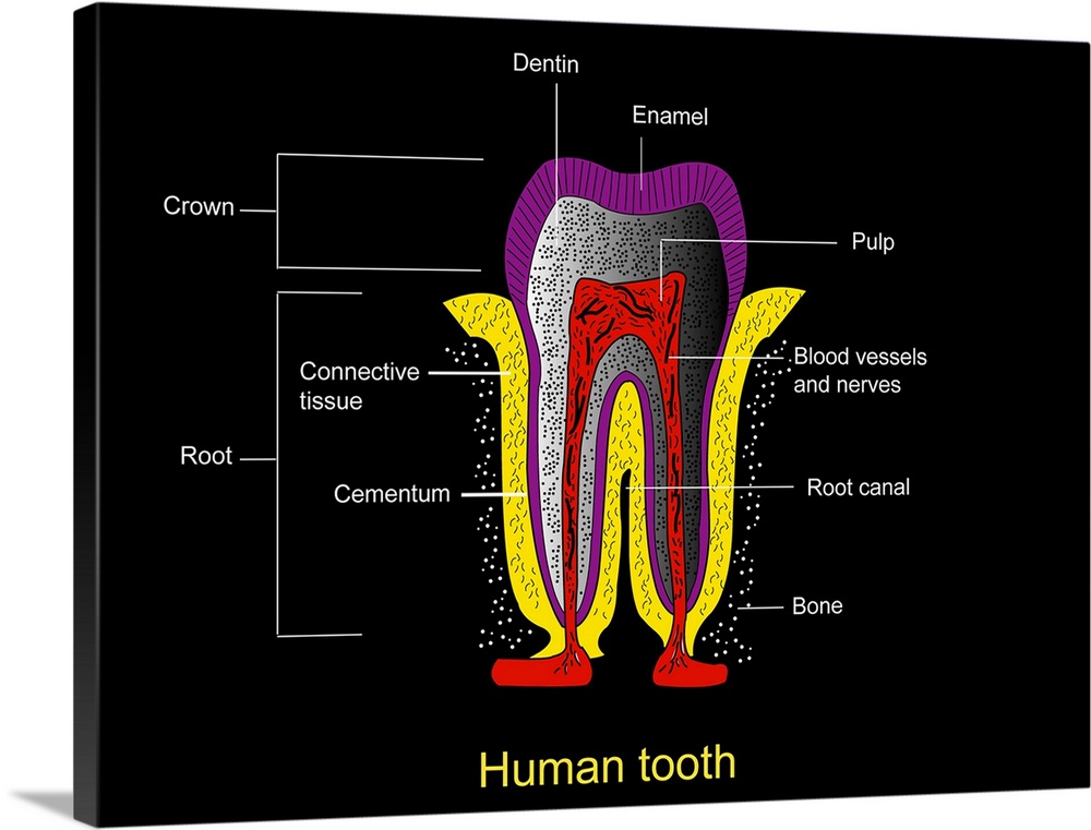
<p>Human tooth anatomy. Diagram of a cross-section through a human tooth to show its anatomical structure. The two main areas are the crown (above the gum-line) and the root (embedded in the gum). The tissue and mineral layers shown here include: enamel (purple, covering the crown), dentin (grey with black dots), the pulp (red), blood vessels and nerves (black, within pulp), cementum (purple, covering the root), connective tissue (yellow), and bone (black with white dots). Also shown is the root canal, the dark space between the two protrusions that form the tooth's root.</p>
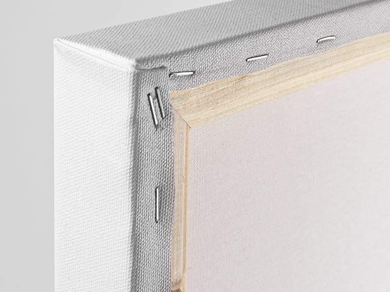
Elevate any room with our handcrafted stretched canvas gallery wraps. Printed with archival inks and wrapped around a 1.25” inch solid wood stretcher bar, our giclée big canvas art prints are a timeless option for any décor style or space.
<p>Our giclée canvas art prints are produced with high quality, UV-resistant, environmentally-friendly, latex inks and artist grade, polycotton canvas. We pride ourselves on color accuracy and image clarity to ensure your new canvas wall art lasts for years to come.</p>

<p>Assembled in the USA, each of our 1.25” inch gallery wrapped canvas art prints is stretched and stapled by our highly skilled craftspeople. Each canvas print is carefully handcrafted to ensure taut canvas wraps and clean corners for outstanding quality and durability.</p>

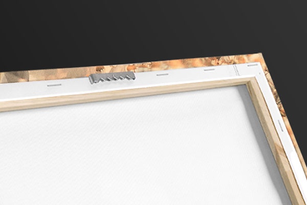
<p>Our handcrafted stretched canvas prints include sawtooth hangers for an easy and secure installation.</p>
 Commercial Decor
Science
Dentistry
Human Anatomy
Illustrations
Francis Leroy
Commercial Decor
Science
Dentistry
Human Anatomy
Illustrations
Francis Leroy