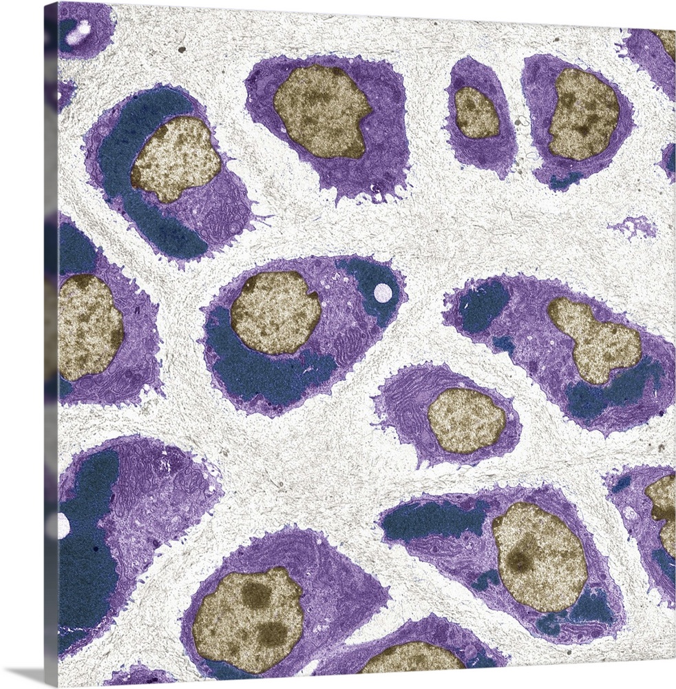
<p>Cartilage cells. Coloured transmission electron micrograph (TEM) of a section through chondrocytes from nasal hyaline cartilage. The cell nuclei (brown) can be seen along with rough endoplasmic reticulum (light purple lines) and glycogen granules (dark purple dots). Chondrocytes are the only cells found in cartilage. Hyaline cartilage is found in the epiphyseal plates of growing bones, and in the nose, larynx (voice box), trachea (windpipe) and bronchus. These cells produce and maintain the cartilage's extracellular matrix. Magnification: x800 when printed 10 centimetres wide.</p>
Elevate any room with our handcrafted stretched canvas gallery wraps. Printed with archival inks and wrapped around a 1.25” inch solid wood stretcher bar, our giclée big canvas art prints are a timeless option for any decor style or space.
<p>Our giclée canvas art prints are produced with high quality, UV-resistant, environmentally-friendly, latex inks and artist grade, polycotton canvas. We pride ourselves on color accuracy and image clarity to ensure your new canvas wall art lasts for years to come.</p>

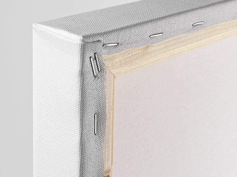
<p>Assembled in the USA, each of our 1.25” inch gallery wrapped canvas art prints is stretched and stapled by our highly skilled craftspeople. Each canvas print is carefully handcrafted to ensure taut canvas wraps and clean corners for outstanding quality and durability.</p>

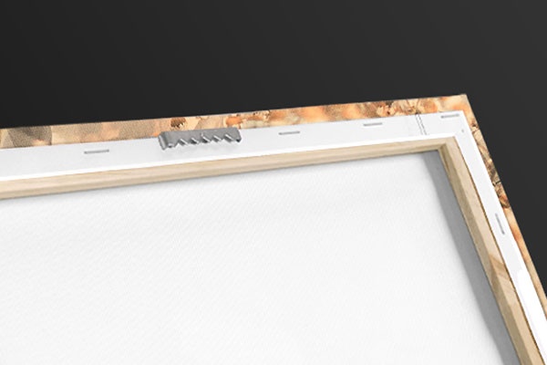
<p>Our handcrafted stretched canvas prints include sawtooth hangers for an easy and secure installation.</p>
 Educational
Science
Biology
Human Anatomy
Steve Gschmeissner
Educational
Science
Biology
Human Anatomy
Steve Gschmeissner