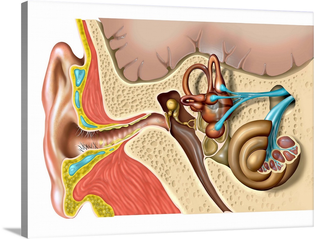
<p>Human ear anatomy. Computer artwork of the structure of the human ear, showing the outer ear (left), middle ear and inner ear (right). The ear drum (tympanic membrane, centre) separates the inner and middle ear, and transmits sound to the ossicles (centre right). This system of tiny bones transmits sound from the air-filled middle ear to the fluid filled cochlea (coiled, lower right) in the inner ear. The cochlea contains the organ of corti (cutaway), which contains rows of hair cells topped with stereocilia. These cilia touch the tectorial membrane and detect tiny movements caused by sound-induced pressures in the inner ear fluids, which are translated into nerve impulses that travel to the brain, where they are deciphered as sound.</p>
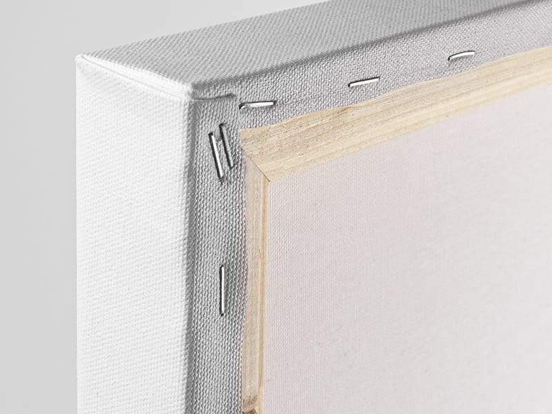
Elevate any room with our handcrafted stretched canvas gallery wraps. Printed with archival inks and wrapped around a 1.25” inch solid wood stretcher bar, our giclée big canvas art prints are a timeless option for any decor style or space.
<p>Our giclée canvas art prints are produced with high quality, UV-resistant, environmentally-friendly, latex inks and artist grade, polycotton canvas. We pride ourselves on color accuracy and image clarity to ensure your new canvas wall art lasts for years to come.</p>

<p>Assembled in the USA, each of our 1.25” inch gallery wrapped canvas art prints is stretched and stapled by our highly skilled craftspeople. Each canvas print is carefully handcrafted to ensure taut canvas wraps and clean corners for outstanding quality and durability.</p>

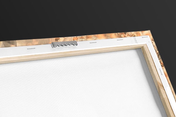
<p>Our handcrafted stretched canvas prints include sawtooth hangers for an easy and secure installation.</p>
 Educational
Science
Human Anatomy
Illustrations
Jose Antonio Peaas
Educational
Science
Human Anatomy
Illustrations
Jose Antonio Peaas