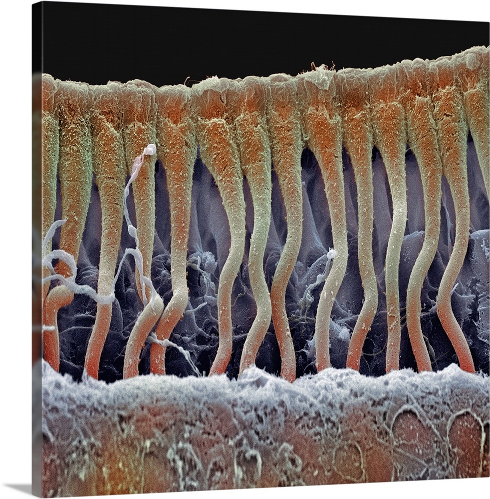
<p>Cochlea cells. Scanning electron micrograph (SEM) of a vertical section through part of the cochlea inside a human ear. The section shows part of the row of columnar outer pillar cells that runs along the organ of Corti, the auditory sense organ. The outer pillar cells arise from the basilar membrane (across bottom), and their upper surfaces (across top) form part of the surface of the organ of Corti. This organ lies on the basilar membrane, an internal surface of the cochlear duct. The organ of Corti also contains hair cells (not seen) and an overlying tectorial membrane (removed). Sound waves deform hair cell cilia and trigger auditory nerve impulses. Magnification: x600 at 6x7cm size.</p>
Elevate any room with our handcrafted stretched canvas gallery wraps. Printed with archival inks and wrapped around a 1.25” inch solid wood stretcher bar, our giclée big canvas art prints are a timeless option for any decor style or space.
<p>Our giclée canvas art prints are produced with high quality, UV-resistant, environmentally-friendly, latex inks and artist grade, polycotton canvas. We pride ourselves on color accuracy and image clarity to ensure your new canvas wall art lasts for years to come.</p>

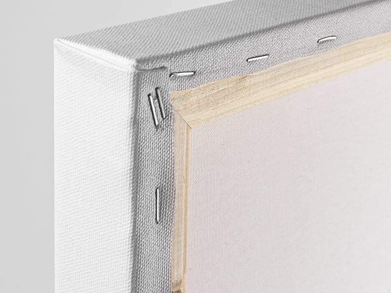
<p>Assembled in the USA, each of our 1.25” inch gallery wrapped canvas art prints is stretched and stapled by our highly skilled craftspeople. Each canvas print is carefully handcrafted to ensure taut canvas wraps and clean corners for outstanding quality and durability.</p>

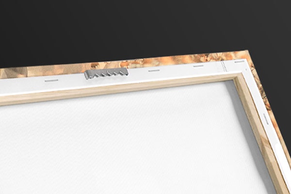
<p>Our handcrafted stretched canvas prints include sawtooth hangers for an easy and secure installation.</p>
 Educational
Science
Human Anatomy
Steve Gschmeissner
Educational
Science
Human Anatomy
Steve Gschmeissner