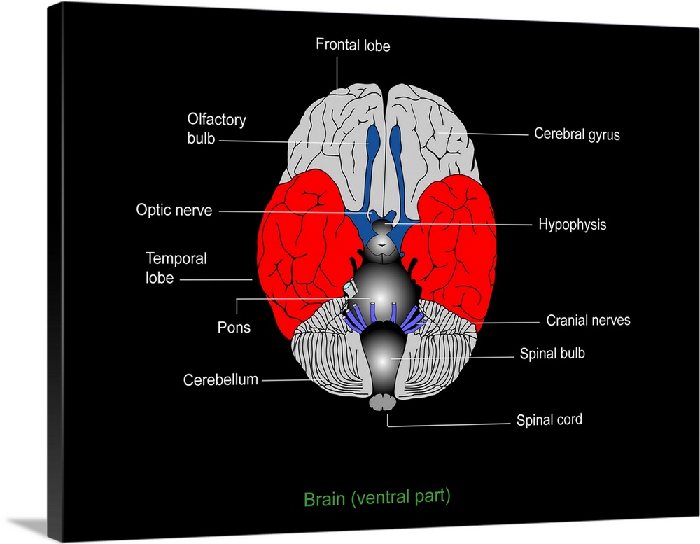
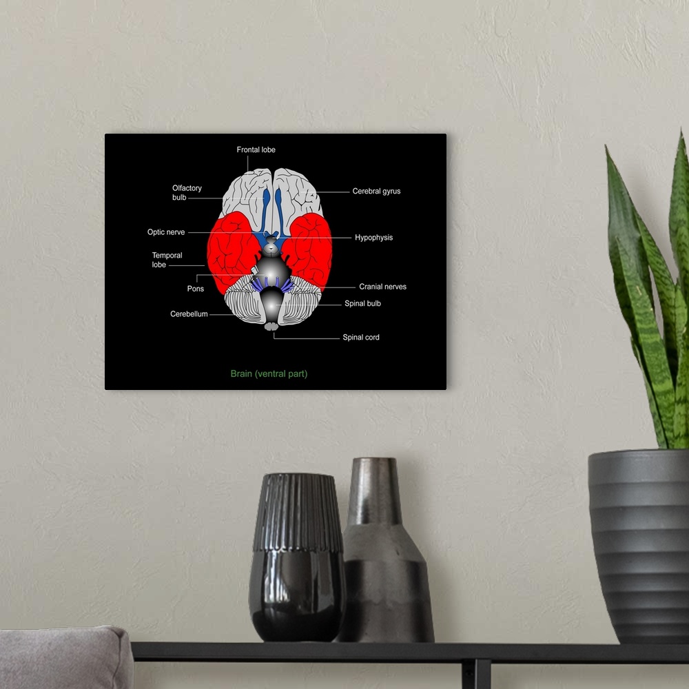
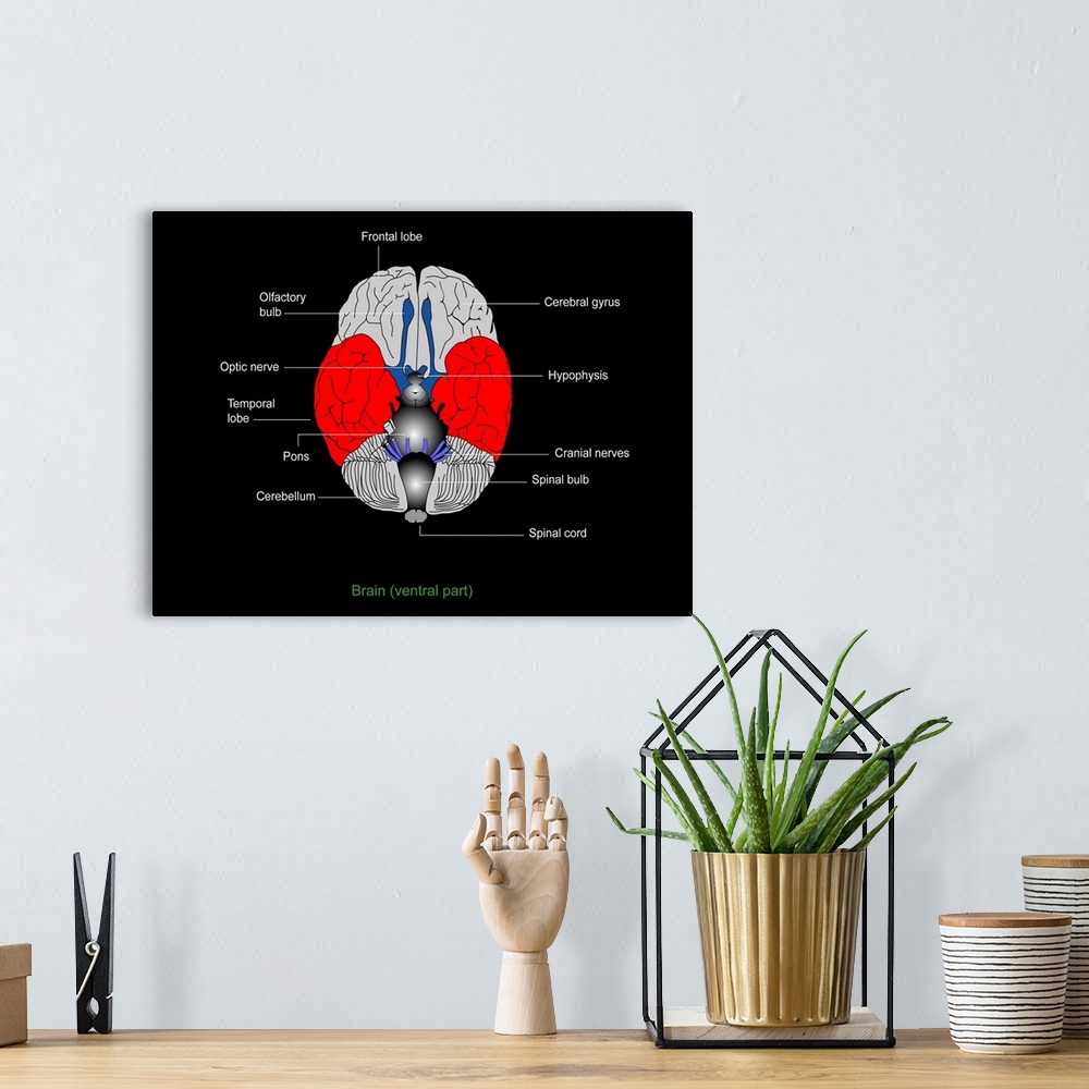
<p>Brain anatomy. Diagram of the underside (ventral aspect) of the brain, showing the anatomical structure of the various components. At the rear of the brain is the cerebellum (grey with black lines), split between the two hemispheres of the brain. Also split between the hemispheres are the temporal lobes (red), and the frontal lobes (top). The folds in the lobes are called gyri. Descending from the frontal part of the brain are the olfactory bulbs and optic nerves (blue, top). Also blue, but lower down, are the cranial nerves. The darker grey areas down centre are (top to bottom) the hypophysis, the pons, and the spinal bulb. Right at bottom is the spinal cord.</p>
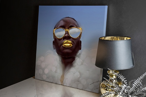
Elevate any room with our handcrafted stretched canvas gallery wraps. Printed with archival inks and wrapped around a 1.25” inch solid wood stretcher bar, our giclée big canvas art prints are a timeless option for any décor style or space.
<p>Our giclée canvas art prints are produced with high quality, UV-resistant, environmentally-friendly, latex inks and artist grade, polycotton canvas. We pride ourselves on color accuracy and image clarity to ensure your new canvas wall art lasts for years to come.</p>
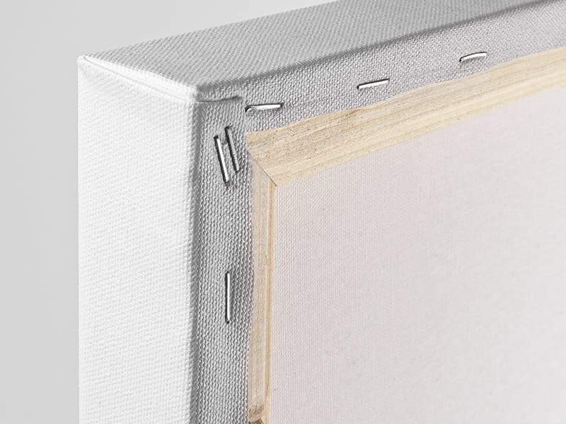
<p>Assembled in the USA, each of our 1.25” inch gallery wrapped canvas art prints is stretched and stapled by our highly skilled craftspeople. Each canvas print is carefully handcrafted to ensure taut canvas wraps and clean corners for outstanding quality and durability.</p>

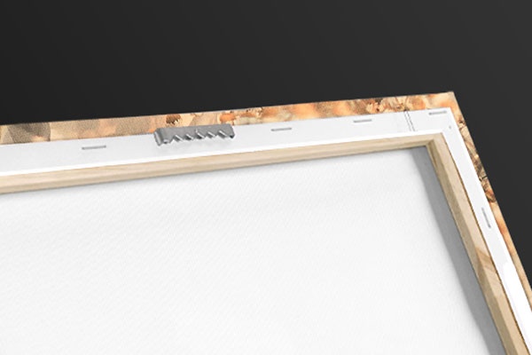
<p>Our handcrafted stretched canvas prints include sawtooth hangers for an easy and secure installation.</p>
 Science
Human Anatomy
Illustrations
Francis Leroy
Science
Human Anatomy
Illustrations
Francis Leroy