{{ titlePrefix }} Biology
About Biology
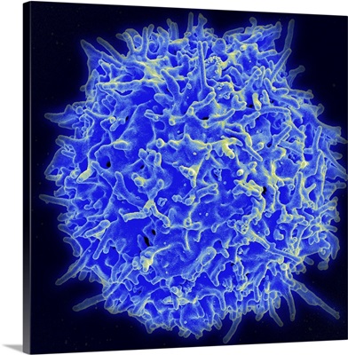
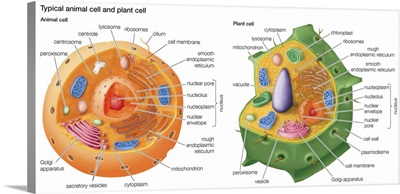
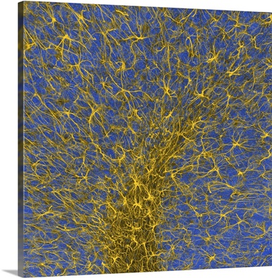
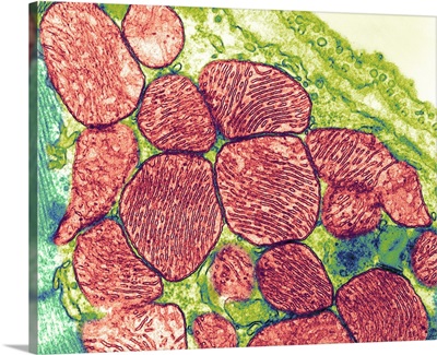
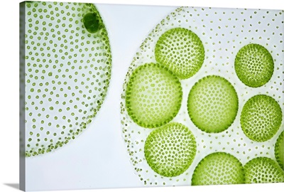
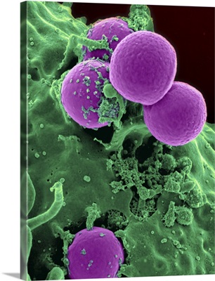
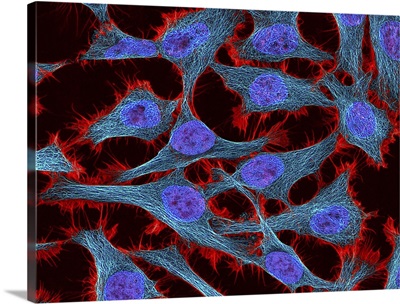
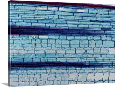
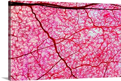
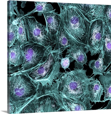
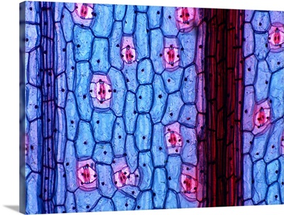
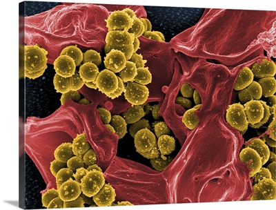
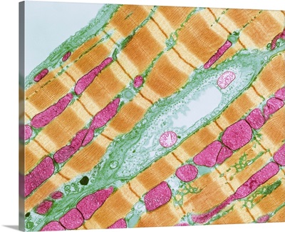
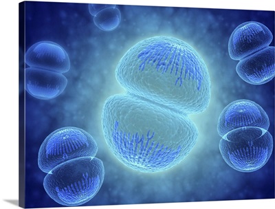
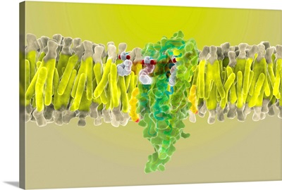
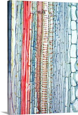
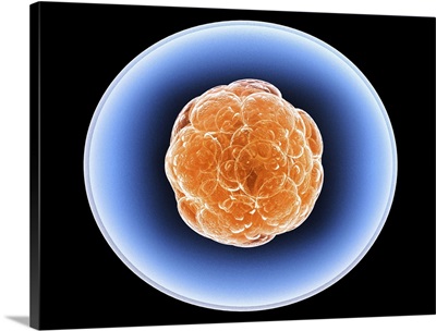
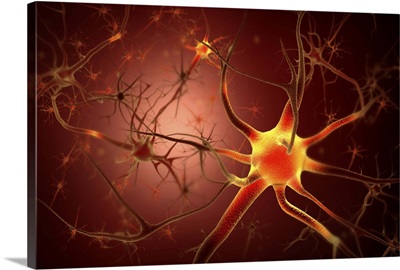
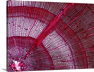
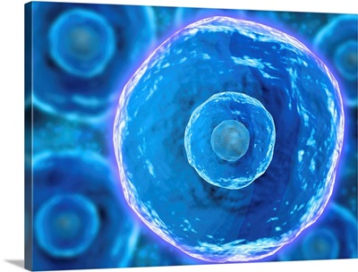
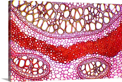
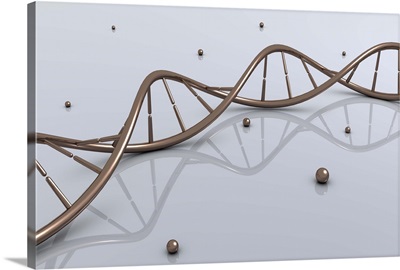
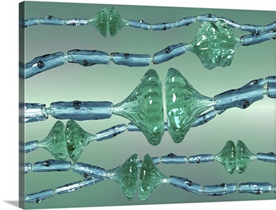
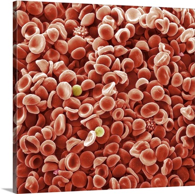
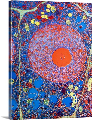
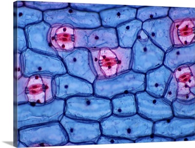
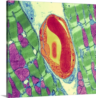
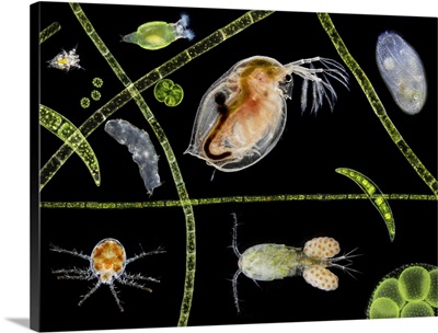
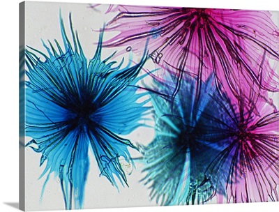
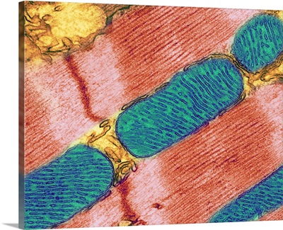
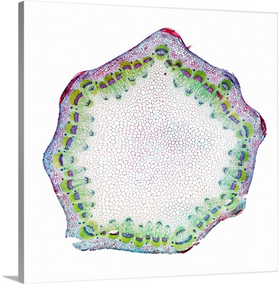
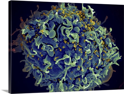
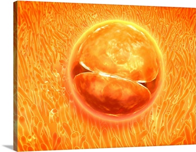
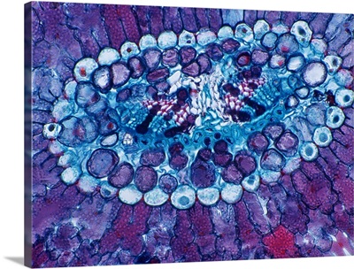
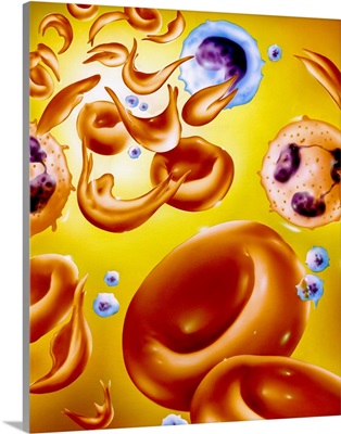
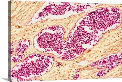
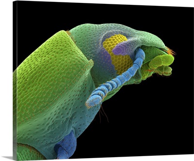
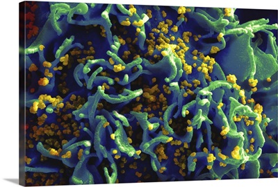
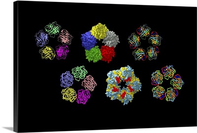
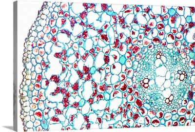
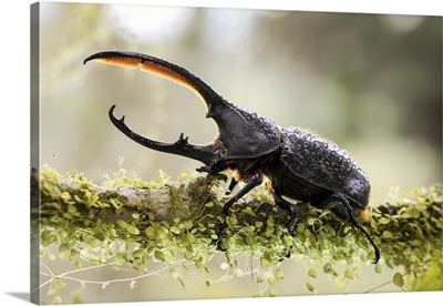
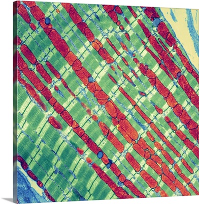
The study of life is a fascinating subject and a uniquely beautiful one, as well! From plant cells to animal cells, mitochondria to DNA, biological images make distinct and appealing art. Magnified prints of microscopic images make gorgeous wall art for the science lover—unique and varied geometric designs are formed by the different types of cells, including blood cells and bacteria. Biology art is perfect for learning or design purposes—images of cells are as appealing as they are educational. Regardless of the use, art inspired by biology offers a captivating glimpse into the smallest fundamentals of life itself